Biochemical measurements
The examinees were instructed to fast for 12 hours and refrain from urination 4 hours prior to examination. Totally 61% of the participants were examined before 12 a’clock, when fasting generally had lasted for over 12 hours. The length of fasting was under 9 hours in 53% of the participants.
Blood determinations: One hour after ingesting glucose solution, given for 1 hour glucose tolerance test, a blood sample was drawn for the determination of plasma glucose. Each person received a 20 % glucose dose graded (250-337 ml, 338-412 ml, 413-450 ml) according to the person’s size/body surface area, which was estimated using a table of height and weight. The glucose solution may have enlarged the plasma volume which may have reduced the concentrations for some of the determinations.
The following tubes were taken:
- an EDTA-tube for hematological (hemoglobin and hematocrit) determinations
- a heparinized tube for plasma glucose and creatinine determinations
- serum tubes for cholesterol, triglycerides, protein bound iodine, iron, TIBC, and uric acid determinations
- one 10 ml serum and plasma tube to be stored in the frozen serum/plasma storage
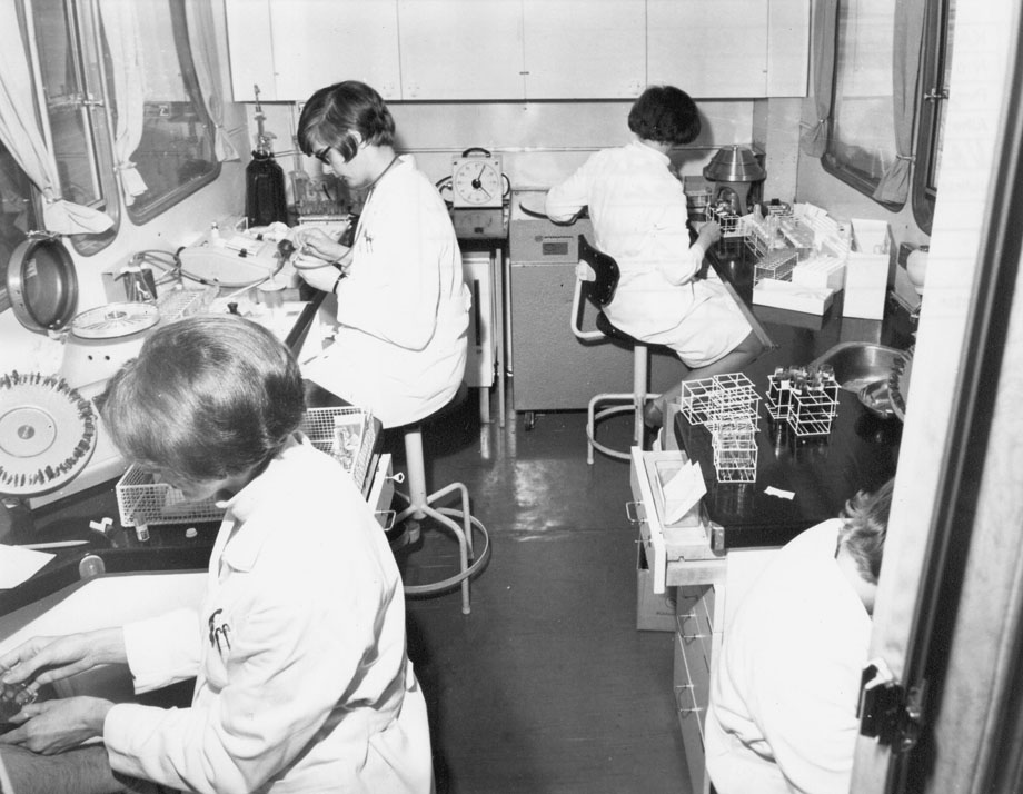
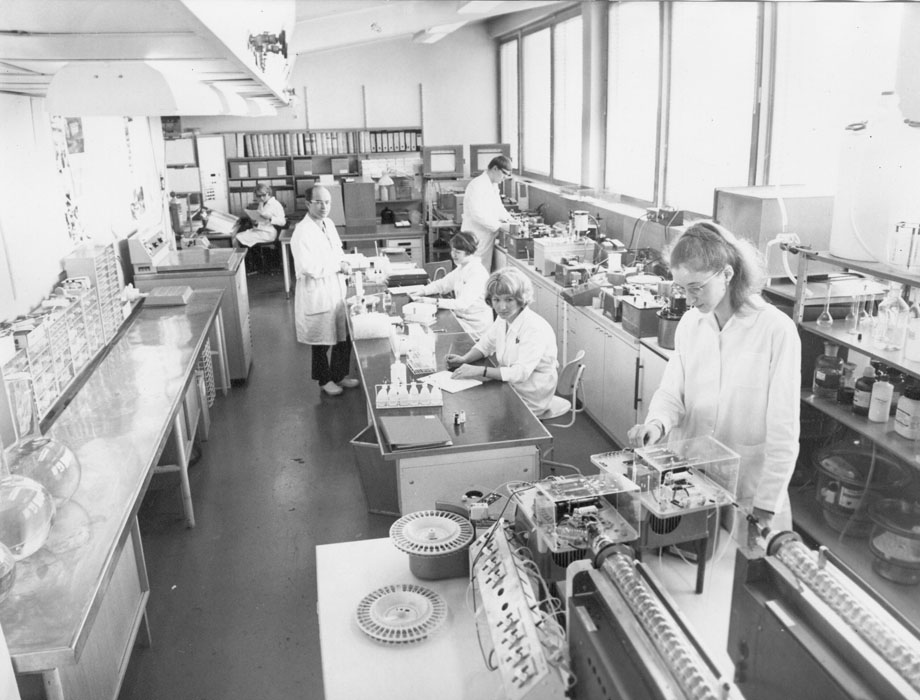
Hemoglobin and hematocrit were determined in the laboratory of the mobile clinic car on the same day the blood sample was taken. The other determinations were made from frozen samples 1−3 weeks later in the Mobile Clinic’s supporting laboratory situated at the National Public Health Institute.
- Hb: Hemoglobin (N = 45 554) was determined using the cyanmethemoglobin method by Vitatron-R-photometry using hemoglobin standard solutions from the Finnish Red Cross Blood Service (Nevanlinna et al. 1964). Also ER (N = 17 828), MCHC (N = 45 553), MCV (N = 17 827), and MCH (17 828) were determined.
- Hkr: Hematocrit (packed cell volume) was based on a blood sample drawn into an EDTA tube and analyzed by the CLAY-ADAMSR microhaematocrit method. The sample was centrifuged for 5 minutes at a speed of 12 500 rpm (Takkunen 1976).
- Cholesterol: Serum cholesterol was determined using an automatic method (Technicon AutoAnalyzer Methodology N-24a 1965, Technicon AutoAnalyzer Methodology N-77 1969, Technicon AutoAnalyzer Methodology 54 1969 ) using the modification of the Liebermann-Burchard method (Huang et al. 1961, Boy 1963). The reliability coefficient (CV) was 0.02 – 0.04.
- Triglycerides: Serum triglycerides (N = 21 727) were determined using an automatic method developed at the Mobil Clinic laboratory (Björksten 1972).
- Glucose: Plasma glucose was determined by the ferricyanide method (Technicon AutoAnalyzer Methodology N-2b, 1965), which on the level 160 mg/100 ml resulted in 5 % higher findings than the enzymatic method. The reliability (CV) was 0.01 – 0.03. In comparison with the other laboratories of the Helsinki control circle the results of the Mobil Clinic central laboratory were 3 – 10 % higher. The results of the US CDC by the enzymatic method were 5 % higher than those analyzed on the same samples by the Mobil Clinic laboratory.
- Creatinine: The creatinine concentration was determined from a plasma sample using a modification of the Jaffe method (Technicon AutoAnalyzer Methodology N-116, 1965). The analytical level of the Mobile Clinic central laboratory was slightly higher than that in the other laboratories of the Helsinki control circle. During the first years, the difference was up to 10 % but since 1970 only a few percentage units. Reliability (CV) was 0.02 – 0.05.
- PSJ: Serum protein bound iodine was determined using an automatic method according to the instruction manual (Technicon AutoAnalyzer Instruction Manual N:o PB/D, 1969). Total iodine was determined for 14 579 participants.
- Iron: Serum iron was determined with a colorimetric method (Technicon AutoAnalyzer Methodology N-62) (Takkunen 1976, Knekt et al. 1994).
- TIBC: Serum iron binding capacity (N = 49 018) was determined using a modification of the automatic Ramsay method (Technicon AutoAnalyzer Methodology N-62, 1968).
- Transferrin saturation: Transferrin saturation (%) was estimated as serum iron/ TIBC.
- Uric acid: Serum uric acid (N = 15 258) was determined using an automatic colorimetric method (Technicon AutoAnalyzer Methodology N-13b).
The serum lipid values of the reference sera (Kliinisten laboratoriotutkimusten laaduntarkkailu Oy) analyzed in the Mobile Clinic central laboratory in 1967-1968 were approximately 10% lower than the means of all laboratories of the control circle. In 1969 they were close to the means of other laboratories. Using control sera the results were also compared with those of the US CDC and the results of the Mobile Clinic central laboratory were about 5 % higher than those of the CDC.
Urinary examination: A clean midstream sample was provided by every examinee. Albumin, glucose and blood were determined qualitatively by test strips. Urinary bacteria were cultured from the women samples. In 1967 and 1968 Drigalski-dishes were used and after that UricultR slides (UricultR). The Drigalski dishes were kept overnight at 37 0 C. Under 20 colonies on the dish were read as zero, 20-90 colonies as 104/ml and over 90 colonies as 105/ml. If growth was over 105 ML the dishes and the slides were sent to the National Public Health Institute for the typing of the bacteria and the determination of antibiotic resistance. The diagnosis bacteriuria was based on two samples each with a growth of 105/ml (Kass 1956, Heinonen et al. 1969).


