Physical measurements
Antropometric measurements: Height was measured without shoes using a scale fixed to the wall. Weight was measured by a heavy duty spring scale, which was calibrated by weights at the beginning of the study in each research place. The examinees were without shoes and they were wearing light indoor clothing. In summer 1 kg and in winter 2 kgs were subtracted from the result. Body mass index was calculated dividing weight by height squared (Heliövaara and Aromaa 1981).
Skinfold thickness (triceps and subscapular, Tanner 1959) was measured by the Harpenden skinfold caliper (British Indicators Ltd, John Bull) with a constant compression of 10 g/mm2 at skinfold thicknesses of 2 – 40 mm.

Blood pressure and the heart rate were measured by an automated instrument employing an aneroid manometer (Elag BPM-A). Based on Korotkoff sounds it registered on paper systolic and diastolic blood pressure, and the heart rate. The size of the rubber bag was 12,5 x 40 cm. The measurement technique (WHO 1962, Rose and Blackburn 1968, WHO 1979, Aromaa 1978) was in accordance with recommendations: The casual blood pressure was measured on the right arm of the sitting subject. The arm rested on the table so that the cuff was at heart level. The pressure was raised 30 mmHg above the systolic blood pressure level estimated based on the pulse. The observer listened to the Korotkoff sounds through a loud speaker and recorded their appearance as systolic and their disappearance (phase V) as diastolic blood pressure. The measurements were carried out by 9 measurers.
The examinee was instructed not to eat, drink or smoke prior to the blood pressure measurement. Blood pressure was measured 10-20 minutes after the ingestion of the glucose dose given for 1 hour glucose tolerance test, which lowered the diastolic blood pressure with about 2 mmHg (Aromaa 1981).
An experiment was carried out on 1 000 persons to estimate how a casual measurement describes the prevalence of permanently raised blood pressure. A threshold value of 160 and 95 mmHg overestimated the prevalence by 2 % and the threshold value of 170 and 100 mmHg by 13 %.

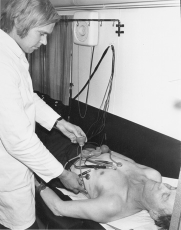
Resting ECG was recorded half an hour after arrival at the examination site, but before the glucose load in 12 municipalities (Mouhijärvi, Vammala, Kauttua, Koskenpää, Jämsänkoski, Jämsä, Rautaruukki, Saloinen, Merijärvi, Joensuu, Kiihtelysvaara, Tohmajärvi) from the 30−59 –years old participants (N = 10 962) (Reunanen 1977, Reunanen et al.1983). The 12-lead resting ECG was recorded by 4 channel Elema-Schönander Mingograf 34-instrument. Paper speed was 50 mm/second. The proper functioning of the galvanometers was checked daily by an ECG simulator (ECG-Simulator EKS-70, Windsor Locks, Connecticut, USA) (Method folder 1.8.1, 1.8.3).
The ECG was interpreted according to the Minnesota code classification system using the revised Minnesota-code (Rose and Blackburn 1968, Scandinavian Committee on ECG Classification 1968, Reunanen et al. 1983). There were some deviations from this code in that ST- and T-wave changes for codes 4.3-4 and 5.3-4 were recorded also when the change was observed only in lead aVF. The ectopic beats were coded according to the criteria of the Scandinavian Committee on ECG Classification (1968).
The ECGs were classified by 6 observers trained and supervised by two cardiologists. The observers worked in groups of 2 or 3. First, every observer independently classified the recordings and their results were compared. If there were discrepancies, the final code was selected by joint decision. The cardiologists checked and classified all ECGs where at least one technician had recorded Q or QS-findings. Every tenth ECG was reclassified to evaluate the repeatability.
Measurement of ECG –items was carried out by 9 specially trained observers supervised by one physician. The physician carried out the measurements of all P- ja T-axes, PTF-V1 and S-T-segments (Ristola ym. 1980, Ristola 1983).
Miniature chest X-ray was taken of everyone except of pregnant women after ingestion of barium paste. The picture size was 100x100 mm. The focal distance was 140 cm. To enhance the evaluation of the pictures a centimeter scale was placed horizontally and vertically in front of the shade plate. The device was Elema - Schönander DAT-154; X-ray tube Siemens PH 125/80; miniature picture camera Old Delft Odelca 100 XVIII; development device Kodak X - Omat M5. Filming values were 120 kV, 150 mA, tube distance 115 cm, focus 1.2 x 1.2 mm, filter 0.7 mm A1. Film was Agfa - Gevaert Scopix T2.
To assess changes of heart size or shape, two radiologists independently classified the pictures using 25 codes of x-ray findings. The between and within variation of the radiologists was rather large, but the agreement with evaluations of large Thorax pictures and the observer’s internal reliability were quite good. The method and the qualities of the evaluation have been described in detail elsewhere (Aromaa et al. 1978). A radiologist evaluated lung changes. Spondylosis and hyperostosis were evaluated and classified by an acquainted internist (Julkunen et al. 1975, 1981, Method folder 1.13).


